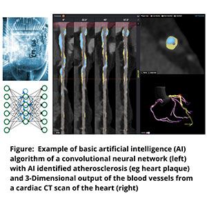
Physicians may be able to quickly and more accurately identify signs for millions at risk of having a heart attack using a new approach that in early stages offers not only provide a better “picture” of someone’s heart, but also uses artificial intelligence (AI) to detect whether someone is potentially at risk of having a heart attack with greater speed and precision.

Those findings were presented as research abstracts by physicians at The George Washington University Medical Faculty Associates (GW MFA), which were awarded as the "Best Abstract Winner" and "1st Runner-Up" out of over 200 international submissions at the Society of Cardiovascular Computed Tomography Scientific Session.
The 15th annual meeting of the Society of Cardiovascular Computed Tomography, held July 17–18, offered a comprehensive exploration of the most current developments in the field of cardiovascular CT principles, methodologies, appropriateness criteria, and clinical practice.
The lead author of the top abstract, Andrew Choi, MD, a cardiologist at the MFA and an associate professor of medicine at the GW School of Medicine and Health Sciences (SMHS), is grateful that his research was chosen, especially because it is the first of its kind.
“Our study, called CLARIFY (CT EvaLuation by ARtificial Intelligence For Atherosclerosis, Stenosis and Vascular MorphologY: A multi-center, international study), was conducted in collaboration with expert medical centers from around the world including Lisbon, Portugal, and southern California. It developed and evaluated a novel artificial intelligence approach that fully automates this complex analysis to, in about 10 minutes, give precision data to the degree, severity, and extent of the complex plaque present in the heart,” Choi explained.

This novel approach is less invasive, and has fewer complications than the traditional practice of inserting catheters into patients to see plaque in the heart’s blood vessels, said Choi. Additionally, a single blood vessel scan can produce up to 100 million voxels (3-D equivalent of a pixel) of data, currently requiring heart imaging specialists to spend up to several hours fully quantifying the plaque present in a single heart scan, Choi noted.
“In the CLARIFY study, we found that when it came to the most severe blockages, the novel AI equaled expert readers for 95–99% percent of the cases,” explained Choi. “We also found that for 1 in 7 cases, AI detected an early blockage that was not readily seen by the expert specialists. When evaluating the presence of plaque by a few millimeters, the AI provided detailed analysis that exceeded the capabilities of the specialists.”
James Earls, MD, a radiologist at GW Hospital and an professor at SMHS, collaborated with Choi on the study. Ultimately, they believe that AI can help cardiologists better look at the whole heart to predict which patients may be at the most risk for a heart attack and also predict a patient’s response to common heart medications, such as statins.
While it’s unlikely that machines will take over a physician’s role anytime soon, AI will continue to integrate with life-saving technologies that are used to make diagnosis of illness and delivery of care more accurate and patient centered.
“Far from science fiction, AI has become an engrained part of society in 2020,” Choi said. “AI can enhance the many challenging ways we diagnose, prevent, and treat heart disease to enable the physician-patient relationship to remain central to outstanding patient care.”


